 |
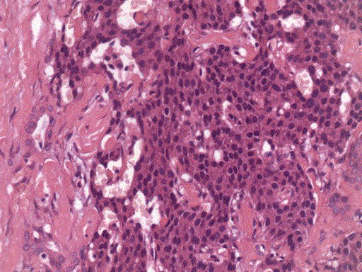
| Figure 3. Histological specimen of insulinoma, (H&E staining, x200 magnification). A trabeculae of epitheloid cells and fibrous stroma, surrounding a central nuclei exhibiting pleomorphism. The cells stained positively for chromogranin, synaptophysin and insulin but negatively for somatostatin, glucagon and gastrin. |