 |
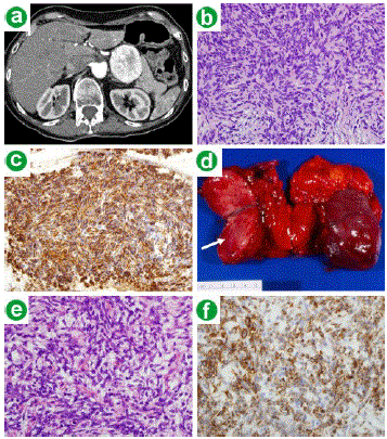
| Figure 1. a. Triphasic CT of the pancreas reveals a 5 cm solid mass arising from the body of the pancreas. b. Cell block H&E stain from the fine needle aspirate specimen showed bland appearing spindle cells (40x). c. Vimentin immunohistochemical stain of the cell block specimen with tumor cells that have strong positivity (40x). d. Pancreatic resection specimen demonstrated a 5.0x5.0x4.5 cm well circumscribed non-encapsulated mass (arrow) present in the body of the pancreas, which is shown bisected. e. H&E stain of the resected tumor revealed spindle cells in a variably collagenous background with minimal cytologic atypia and rare mitotic activity (40x). f. Bcl-2 immunohistochemical stain of the resected mass with tumor cells that have strong positivity (40x). |