|
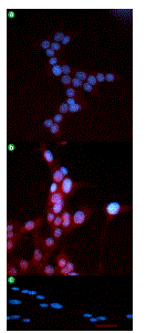
| Figure 3. Immunocytochemical localization of angiotensin II type 1 receptor (AT1R) in INS-1E betacells. Superimposed 4',6'-diamidino-2-phenylindole (DAPI) (nuclei; blue) and rhodamine (AT1R; red) images revealed that AT1R-labeling was detected in cells treated with 5.6 mM glucose (a.) and with 28 mM glucose (b.). c. Negative control image with the omission of primary antibodies. (Magnification: 63x; scale bar: 40 μm). |