|
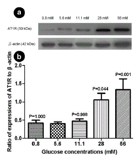
| Figure 2. Western blot analysis of the angiotensin II type 1 receptor (AT1R) protein from INS-1E beta-cell lysates after incubation with a range of glucose concentrations. a. Representative gel image. A major band of about 50 kDa was detected. Control beta-actin expression is shown. b. Expression of AT1R protein relative to beta-actin. All data are expressed as mean±SE; n=5 per group; P values vs. 5.6 mM glucose: ANOVA followed by Tukey’s post hoc test. |