|
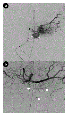
| Figure 3. a. Angiogram performed with catheter tip in origin of dorsal pancreatic artery (black arrow) demonstrating pseudoaneurysm of transverse pancreatic artery (white arrows). b. Coeliac axis angiogram post embolization demonstrating pseudoaneurysm to be excluded from the circulation by multiple platinum coils (white arrows). |