|
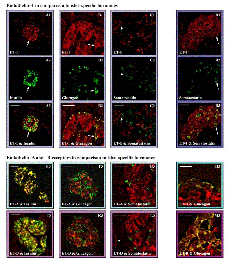
| Figure 1. Expression of endothelin-1 (ET-1) and its corresponding receptors ETA and ETB in comparison to isletspecific hormones in human and rat pancreatic sections. Positive staining for components of the endothelin system are shown in red whereas islet specific hormones are visualized in green. The yellow color in the merged pictures (A3-M3) represents the co-expression of indicated antigens within individual cells. Arrows designate identical cells within the corresponding column. Rat sections are only shown where immunofluorescence staining differs from human sections. L3, arrow head: positive immunostaining of ETB-receptor in vascular endothelium. Bars = 50 μm. |