|
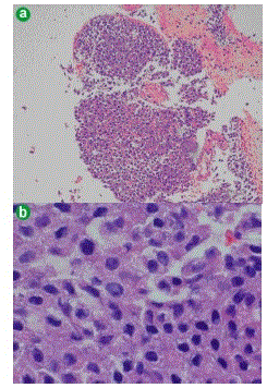
| Figure 1. Light microscopic appearance of acinar cell carcinoma. a. At low-power microscopy, the tumor was hypercellular, with cords of cytologically uniform cells with granular eosinophilic apical cytoplasm reflecting the accumulation of zymogen granules. On cell block, the tumor cells arranged in thick trabeculae, poorly formed glands or acini. (H&E stain x4). b. At higher power microscopy, an abundant pink, eosinophilic cytoplasm, with granules was visible. Tumor cells with pleomorphic, centrally located nuclei, small to prominent nucleoli and scant to moderate amount of cytoplasm. Brisk and abnormal mitosis are evident. (H&E stain x40). |