|
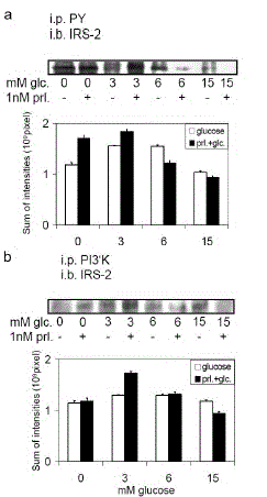
| Figure 5. Prolactin increases IRS-2 activation mainly in the presence of low glucose concentrations. INS-1 cells (80% confluent on a 15-cm diameter dish) were stimulated with 0. 3, 6 or 15 mM glucose plus/minus 1 nM prolactin for 10 min. Cell lysates were subjected to immunoprecipitation (i.p.) with antiserum against phosphotyrosine residues (PY) (a.) or against the p85 regulatory subunit of PI3‘K (b.). Immunoprecipitates were then subjected to immunoblot (i.b.) analysis with anti-IRS-2 antibodies. Experiments were done at least three times. A representative blot for coimmunoprecipitation analysis is shown. Quantification was performed by densitometric scanning with Bio- 1D® (Vilber Lourmat, Eberhardzell, Garmany) software analysis and is presented as the sum of intensities (109 pixel). |