 |
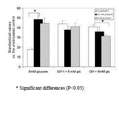
| Figure 3. [3H]thymidine incorporation in INS-1 cells stimulated with 0.5 or 2nM prolactin with or without additional growth factors. Approximately 105 quiescent INS-1 cells/well were incubated for 24 h in RPMI 1640 medium containing 0.1% BSA, 6 mM glucose plus/minus 0.5 or 2 nM prolactin plus/minus 10 nM IGF-1 plus/minus 10 nM GH, then assessed for proliferation rate by [3H]thymidine incorporation. All experiments were done in triplicate on at least five independent occasions. Data are presented as x-fold standardized value above the control observation in the absence of glucose and prolactin, and depicted as a mean±SE (n=5). * Significant differences (P< 0.05) |