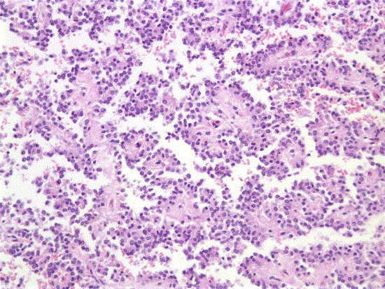
Figure 5.
Microscopic appearance of SPT (Case 2): characteristic pseudopapillary growth pattern with gaplike spaces and delicate fibrovascular stalks lined by a single row of tumour cells (H&E stain x100).