 |
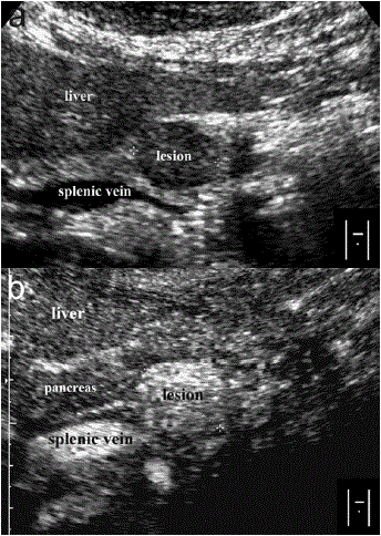
| Figure 2: Pancreatic metastsis of a renal cell carcinoma at conventional (a.) and echo-enhanced sonography (b.). a. Low-echo mass with sharply delinated margins without dilatation of Wirsung's duct. b. Highly vascularized lesion compared to the surrounding tissue. |