|
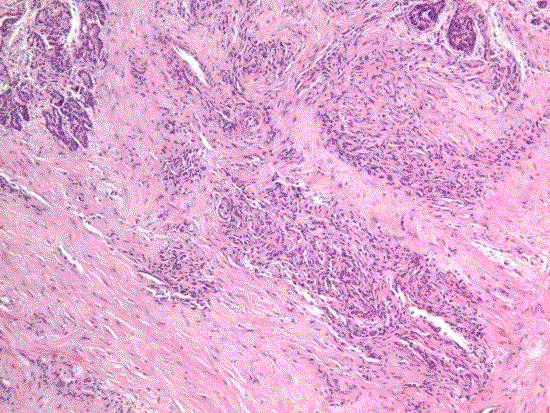
| Figure 4. The biopsy shows lobules of small capillary vascular channels separated by densely sclerotic stroma replacing the pancreatic tissue. Some residual acinar pancreatic tissue is seen in the upper right of the specimen. (Hematoxylin and eosin, x100). |