|
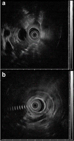
| Figure 4. Endoscopic ultrasound. a. Ventral pancreas and main pancreatic duct having a normal caliber, increased duct wall echogenicity and a cystic lesion (1.6x2.2 cm). b. Absence of the accessory pancreatic duct and the dorsal pancreas. The splenic vessels are in contact with the posterior wall of the stomach. |