 |
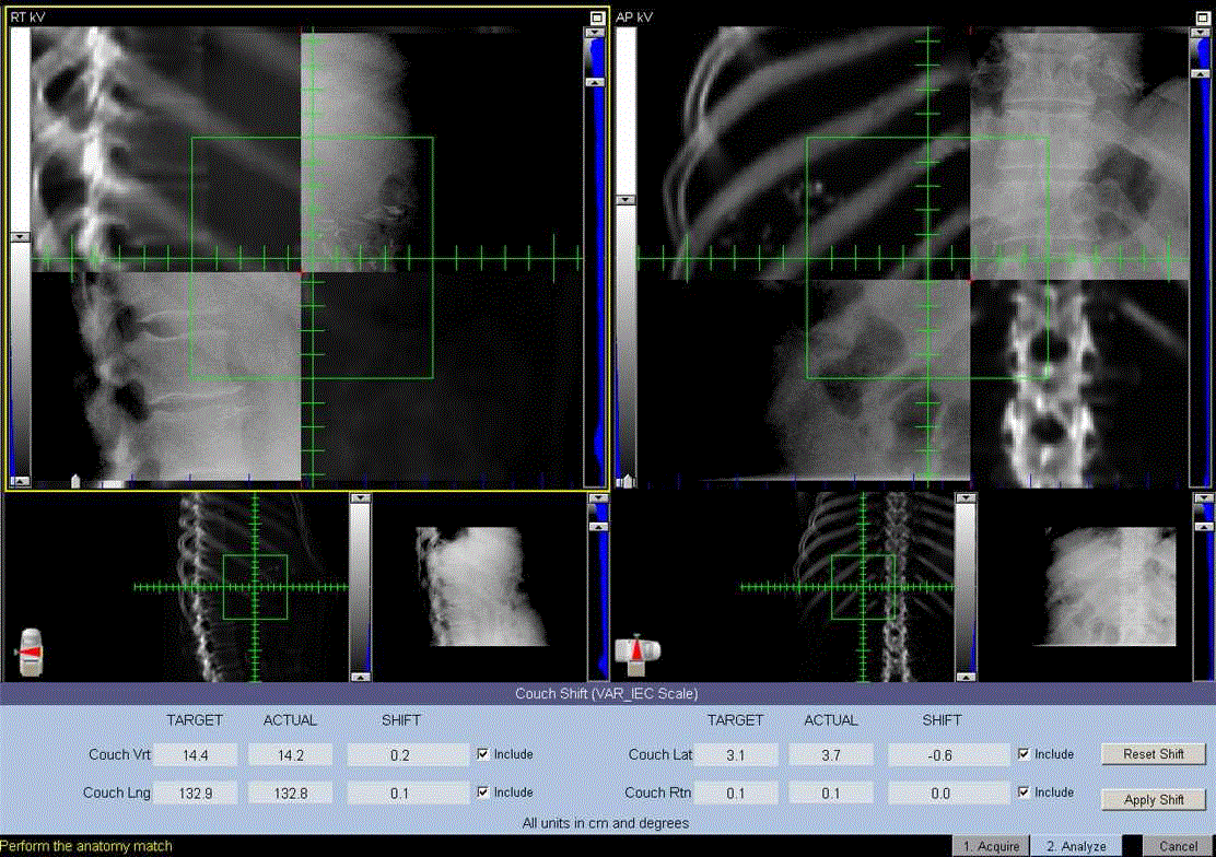
| Figure 1. Composite image of a kilovoltage (kV) image (upper right and lower left boxes) obtained on the day of treatment with the digitally reconstructed radiograph (DRR); upper left and lower right) from the CT image at the time of initial simulation. Image matching to bony landmarks or radiopaque surgical clips were made automatically or manually. In this case, the software automatically calculated a shift of 0.2 cm vertically (Vrt), 0.1 cm longitudinally (Lng), -0.6 cm laterally (Lat), and 0.0 cm rotationally (Rtn). |