|
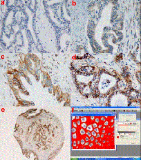
| Figure 1. Digital images of different HER2/neu protein expression cases. a. score 0; b. score 1+; c. and d. scores 2+ and 3+, according to conventional eye microscopy: image analysis identified borderline, but distinct levels of optical density and, finally, those cases which were characterized as 2+ (c.) and 3+ (d.), respectively (original magnification: 40x); e. a tissue microarray spot (core diameter: 1 mm) stained by HER2/neu monoclonal antibody TAB 250 (original magnification: 2.5x); f. image analysis process (red areas represent the values of HER2/neu membranous immunostaining). |