|
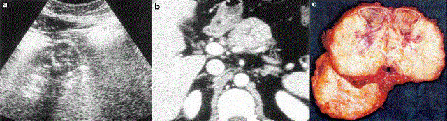
| Figure 4. Case 4. US revealing a round, well-defined, non-homogeneous mass of the pancreatic body, 5x4 cm in diameter, with a thin capsule and multiple central foci of calcifications (a.). A computed tomography scan confirmed the ultrasound findings (b.). A central pancreatectomy was performed and the mass appeared as a solid and cystic lesion (c.). (Image c. is presented in another contribution by the same authors, published in these Proceedings [30], in order to describe aspects not related to those reported here) |