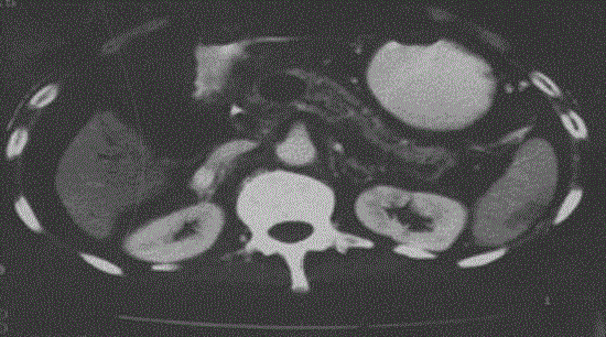
Figure 2.
Post radiofrequency ablation contrast enhanced computed tomography demonstrating 3 cm diameter necrosed (non enhancing) area.