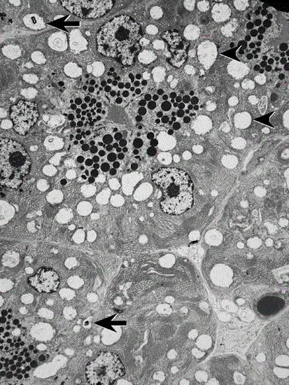
Figure 3.
Electron microscopy , 2000 magnification, of Dog D pancreas showing pancreatic cellular injury as demonstrated by the presence of myelin figures (arrows) and swollen mitochondria with loss of christae (arrowheads).