 |
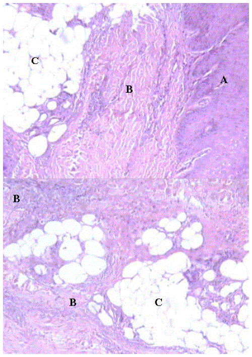
| Figure 3. Septal panniculitis: histopathological microphotograph. A low power microscopic view shows a normal epidermis (A) with lymphoplasmacytic infiltration along the fibrous septa (B) in between the subcutaneous fat lobules (C) and around the dermal blood vessels suggestive of a very early stage of pancreatic panniculitis. |