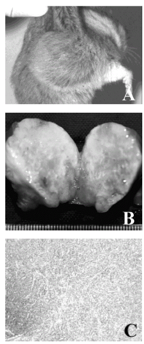
Figure 2:
A case from the SCI group. A. Appearance of the right lymph node. B. Necropsied lymph node specimen cut sagitally. C. Histopathologic view of the lymph node (H&E, 100x).