 |
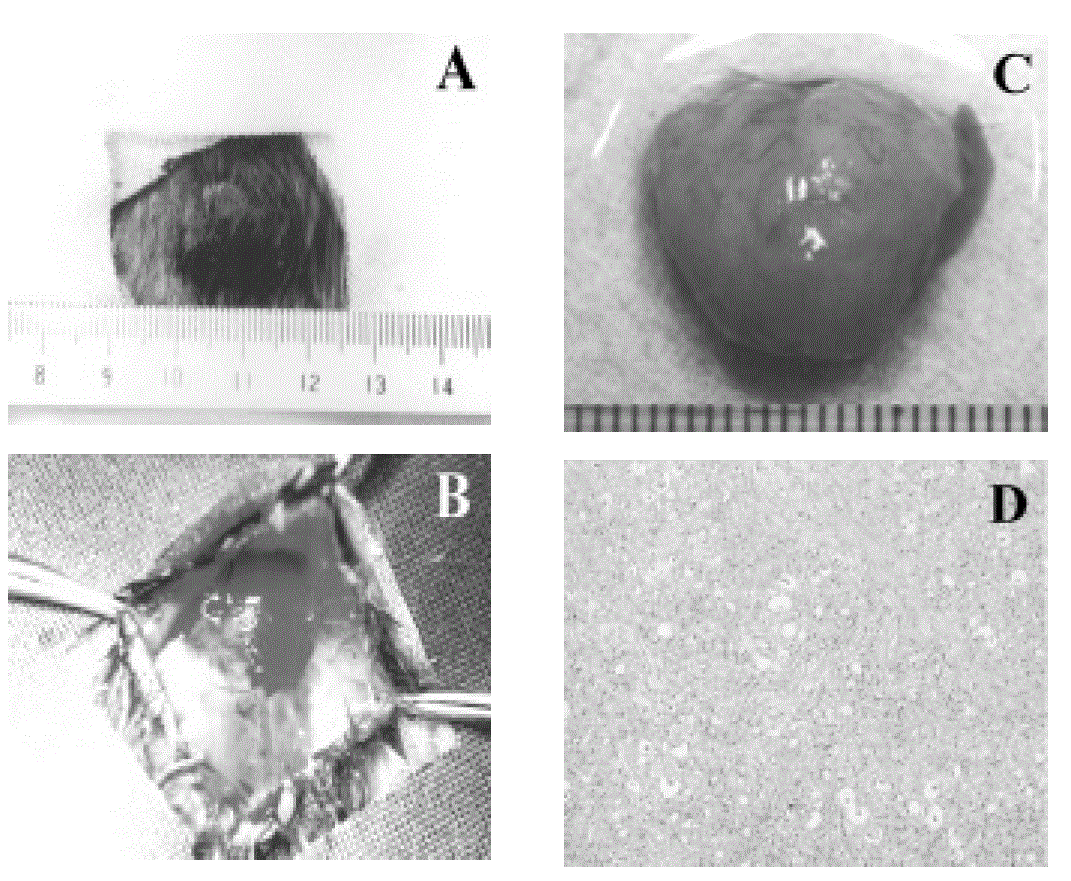
| Figure 1: A subcutaneously implanted hamster. A. Appearance before tumor resection. B. Panoramic view after resection. C. Resected specimen without the covering skin. D. Histopathologic view showing a moderately differentiated adenocarcinoma (H&E, 200x). |