|
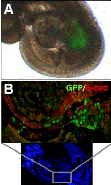
| Figure 2. Gene targeting of a restricted region of the mouse endoderm by whole embryo electroporation. A. Overlay of bright field and fluorescence images of an embryo electroporated at the 8-somite stage with a GFP expression vector and cultured for 24 h, showing GFP activity (green) only in the midgut region. B. Immunolocalization of GFP (green) in the E-cadherin (red)-positive epithelium of the primitive gut. The GFP positive region is boxed in the lower panel, which shows Hoechst staining of a section of the whole embryo. |