 |
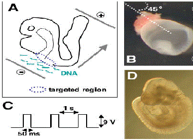
| Figure 1. Electroporation and culture of e8.5 mouse embryos. A. The embryo (drawn here without the visceral yolk sac and amnion) in the electroporation cuvette between the cathode and the anode to target the gut endoderm. The movement of the negatively charged DNA molecules (green) is indicated by the arrow. The region targeted by the DNA is shown in stippled blue. B. Picture and orientation of the embryo (8-somite stage) in the electroporation cuvette prior to electric pulses. C. The square wave pulses used for electroporation of e8.5 embryos. D. Development of an embryo electroporated at the 8-somite stage and cultured for 24 h. Note the progress in development as compared to the embryo shown in panel B. |