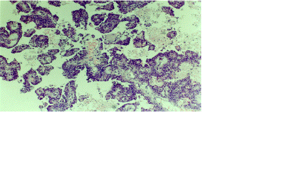
Figure 3.
A micrograph showing cystic degeneration with solid and pseudopapillary formations and red blood cells in the cystic space. (H&E stain. x100).