| i |
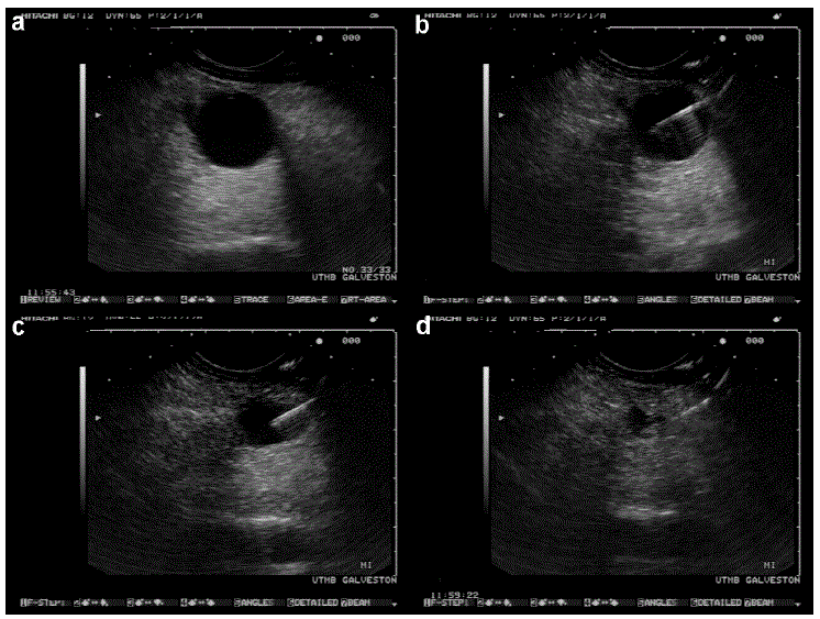
| Figure 1 a. Unilocular cystic lesion in pancreatic body as imaged with a linear echoendoscope in a woman with no symptoms attributable to this lesion. b. Linear EUS guided puncture of the cystic pancreatic lesion shown in Figure 1a. c, d. Progressive decrease in the size of the cystic lesion as cyst fluid is aspirated. Cytopathology revealed mucinous cyst adenoma. |