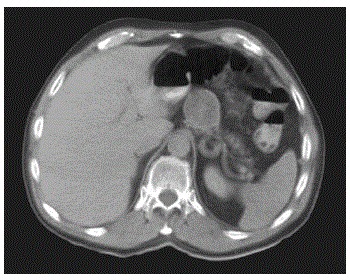
Figure 2.
CT scan performed with intravenous and oral contrast shows a thin walled cystic mass extending anterior from the body of the pancreas. The internal septations are not seen on this scan.