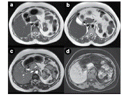
Figure 2.
Axial T2 weighted MRI images (a., b.), TRUFI image (c.) and T1 fat sat image (d.) showing normal head and uncinate process of the pancreas with non-visualization of neck, bodyand tail.