|
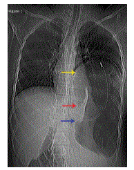
| Figure 1. Scanogram reveals a markedly distended stomach (yellow arrow) with Ryle’s tube in situ. There is an inverse relation of the gastrooesophageal junction (blue arrow) and the pylorus (red arrow)- a finding consistent with intrathoracic gastric volvulus |