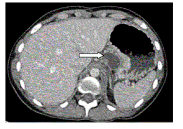
Figure 3.
Case #2. Axial CT image of the patient in figure 1 acquired in portal phase following intravenous contrast medium. This demonstrates the component of the pseudocyst abutting the pancreas (arrow).