|
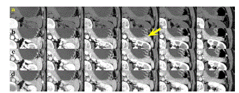
| Figure 1. Axial CT scan of patient 1 (slice thickness: 3 mm) at 0 seconds (pre-contrast) (a), 35 seconds (b), 55 seconds (c), and 80 seconds (d) after the administration of contrast agent, respectively. Although images of (a), (c), and (d) show no remarkable findings, only one slice in series (b) shows a wellenhanced small mass in the tail of the pancreas (indicated by a yellow arrow). |