|
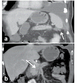
| Figure 1. (a) Reformatted coronal CT image shows a small volume of rim enhancing fluid in the left upper quadrant tracking from the tail of the pancreas to the left hemidiaphragm (arrow). A large left pleural effusion is also seen. (b) Coronal CT image shows dilatation of the main pancreatic duct in the body of the gland with an abrupt cut-off in the region of the pancreatic head at the site of a 2.3 cm mass (arrow). |