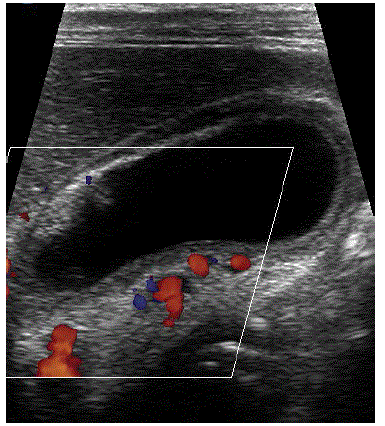
Figure 3:
Sonogram depicts dilated venous channels in the wall of gall bladder.