|
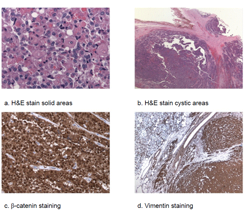
| Figure 1. Histopathology and immunohistochemistry of SPT. a. Hematoxylin and eosin (H&E) stain of solid tumor areas demonstrating uniform polygonal cells with eosinophilic neoplasm, round nuclei with finely stippled chromatin and frequent groves admixed within capillaries. b. H&E stain of cystic tumor areas demonstrating mixed cysts characterized by degenerative changes with formation of pseudopapillary structures. c. Immunoreactivity for β-catenin shows cytoplasmic and nuclei positivity in most tumor cells. d. Vimentin staining is positive in almost all tumor cells. |