|
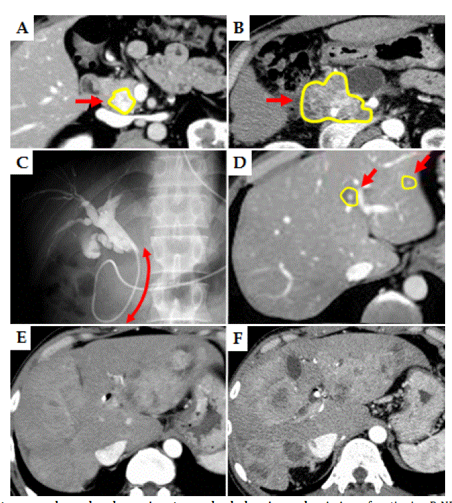
| Figure 1. Enhanced computed tomography and endoscopic retrograde cholangiography. A: A nonfunctioning P-NET in the pancreatic head (2.5 cm in diameter) was observed (yellow circle and red arrow); B: Primary tumor grew, even after therapy (yellow circle and red arrow); C: Obstruction of bile duct due to enlarged primary tumor (red arrow) at 3.5 years after initial diagnosis; D: Small but multiple liver metastases were detected at initial diagnosis (yellow circle and red arrow). E; Liver metastases temporarily seemed to be stable, although continuous and repeated therapies (TACE/TAI, systemic chemotherapies and biotherapies) were required; F: Liver metastases enlarged again at 3.5 years after initial diagnosis. |