|
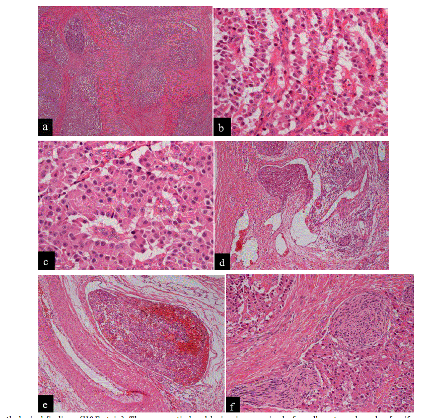
| Figure 3. Histopathological findings (H&E stain). The pancreatic head lesion is comprised of small nests and cords of uniform cells (a.) arranged in a trabecular or ribbon-like pattern (b.). The pseudo-rosette formation (c.) is seen and the intratumoralstroma has rich fibrosis. Lymphatic (d.), venous (e.) and neural invasions (f.) are observed in the pancreatic head region. |