 |
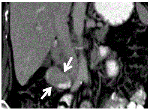
| Figure 1. Computed tomography obtained with oral and intravenous contrast material: coronal reformation image. An enhancing, well-marginated 5.5-cm soft-tissue intraluminal mass (arrows) is seen in the second portion of the duodenum extending to the ampullary region without obstruction. |