 |
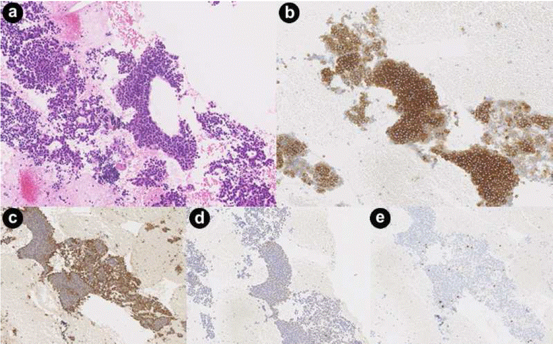
| Figure 3. Examination of the biopsy specimen obtained by endoscopic ultrasound-guided fine needle aspiration showed a proliferation of almost uniform polygonal cells with hyperchromatic round nuclei and eosinophilic fine granular cytoplasm, arranged in alveolar nests, sheets, or partial rosette-like patterns (a.). Immunohistochemical staining showed that many tumor cells were positive for somatostatin (b.), synaptophysin (c.), and chromogranin A (d.). The MIB-1 labeling index was less than 5% (e.). |