|
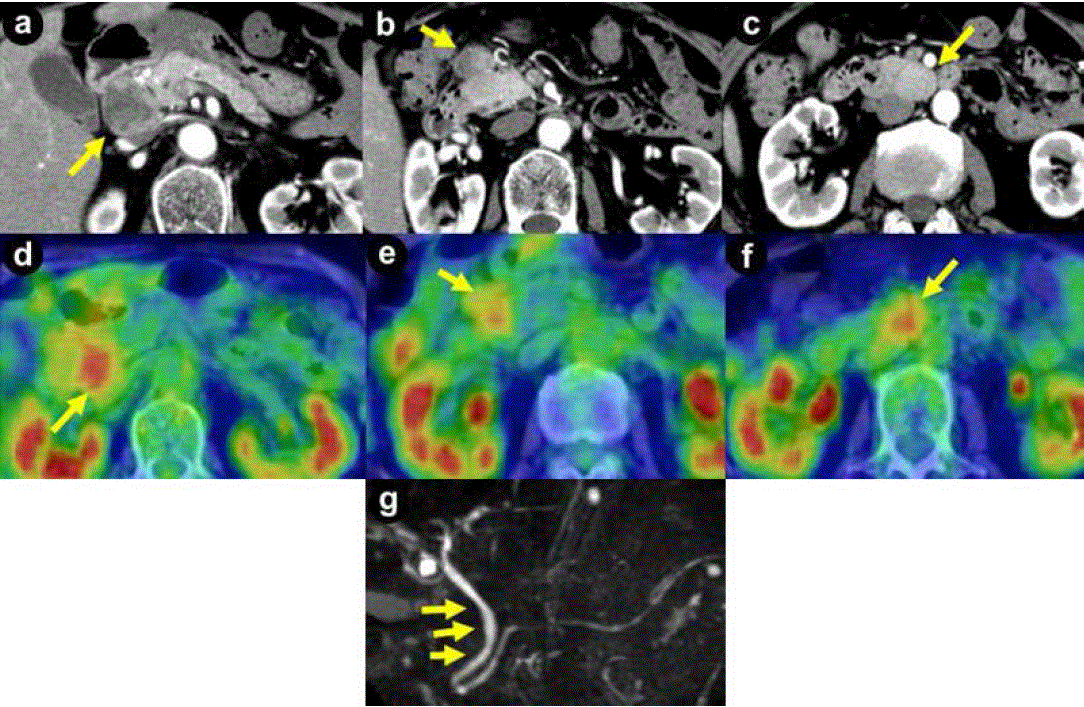
| Figure 1. Enhanced computed tomography showed a low-density mass measuring 37 mm in diameter in the head of the pancreas (a.) and enlarged lymph nodes on the anterior surface of the pancreatic head (b.) and the posterior surface of the horizontal part of the duodenum (c.), measuring 28 mm and 34 mm in diameter, respectively. 18F-fluorodeoxy-glucose positron emission tomography showed increased uptake in all three lesions, with a maximum standardized uptake value ranging 3–4 (d. e. and f.). Magnetic resonance cholangiopancreatography showed that the middle portion of the common bile duct was shifted to the left by the tumor in the head of the pancreas (g.). |