 |
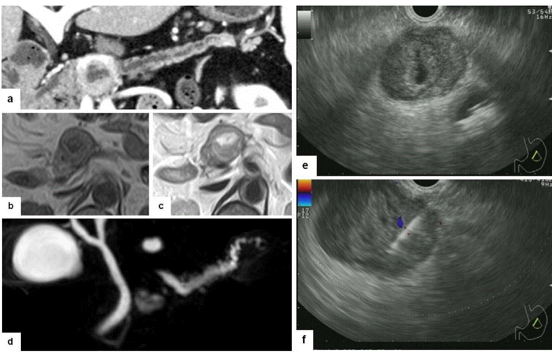
| Figure 1. Findings of image examinations. Curved planar reformation of computed tomography made by tracing the main pancreatic duct showed a well-defined mass of the pancreas with strong enhancement, stenosis of the main pancreatic duct, and central cystic change (a.). Magnetic resonance imaging showed an iso-intensity mass with a central cystic portion and capsule like rim on (b., c.). MR cholangiopancreatography clearly showed cystic degeneration in the central portion of the mass and pancreatic duct stenosis due to the mass (d.). Endoscopic ultrasound demonstrated a well-demarcated hypoechoic mass with a central hyperechoic area, which indicated intratumoral hemorrhage or degeneration (e.). Fine needle aspiration was performed using a 22-gauge needle (f.). |