 |
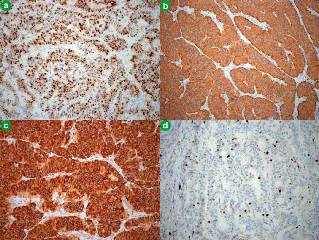
| Figure 3. Immunohistochemical examination of the resected pancreatic mass (magnification power, 40x). a. Tumor cells stained positive for chromogranin A. b. Tumor cells stained positive for synaptophysin. c. Tumor cells stained positive for insulin. d. Tumor cells stained positive for Ki-67 (6%). |