|
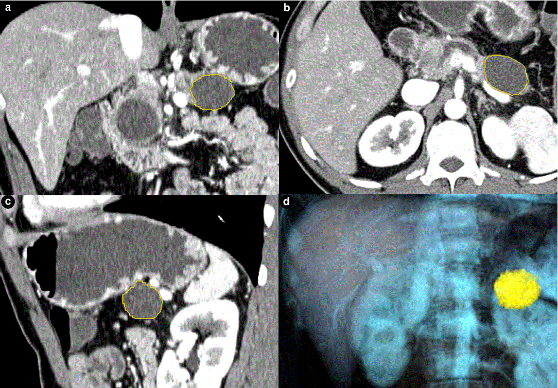
| Figure 2. Coronal (a.), axial (b.), sagittal (c.) and three dimensional (d.) CT images of a histopathologically proven pancreatic tail mucinous cystic neoplasm in a 39-year-old man. Despite a dilated pancreatic duct no connection was visualized on endoscopic ultrasound with the cyst. Borders of cyst are marked with yellow line by the software. CT volumetry (22.3 mL) and an elongation value (0.67) for the cyst were automatically generated by the software once the measurement was finalized by the observer. Pancreatic head cyst was not segmented because of apparent connection with the pancreatic duct that was dilated. Also, note that the patient was a male with mucinous cystic neoplasm, an extremely rare occurrence. |