|
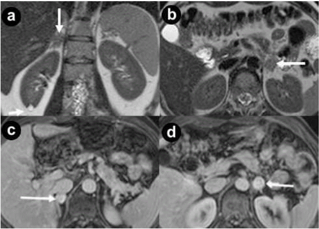
| Figure 8. Adrenal pheochromocytomas. A 38-year-old man with VHL disease. Coronal (a.) and axial (b.) T2-weighted MR images; axial (c. d.) 3D volumetric gradient-echo T1-weighted fat suppressed images after intravenous contrast medium administration during portal venous phase of contrastographic dynamic study. In both adrenal glands, small, round solid masses (arrows), with maximum diameter of 20 mm and intermediate signal intensity in T2- weighted MR images (a. b.) are present. The nodules show intense and homogeneous enhancement after intravenous contrast medium administration during dynamic study (c. d.). Small cyst at lower pole of right kidney (a. short arrow). Bilateral open adrenalectomy was performed. Histological specimen showed the presence of adrenal bilateral pheochromocytomas. |