|
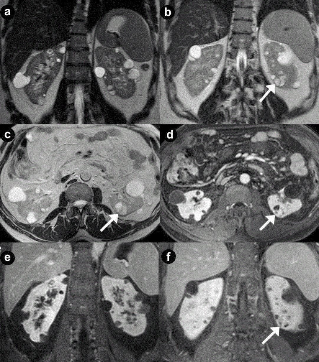
| Figure 7. Renal cysts and cystic renal cell carcinoma. A 43-year-old man with VHL disease. Coronal (a. b.) and axial (c.) T2-weighted MR images; axial (d.) and coronal (e. f.) 3D volumetric gradient-echo T1-weighted fat suppressed images after intravenous contrast medium administration during arterial (d. e.) and venous (f.) phases of contrastographic dynamic study. Multiple and bilateral simple, fluid renal cysts, with high signal intensity on the T2-weighted images (a. b. c.) and hypointense without enhancement after intravenous contrast medium administration during arterial (d. e.) and venous (f.) phases. In the lower pole of the left kidney a complex cystic mass (arrows) is present, with heterogeneous hyperintensity on the T2-weighted images, septa and solid areas enhanced after intravenous contrast medium administration during arterial (d. e.) and venous (f.) phases. Surgical enucleation of left renal lesion was performed. Histological specimen showed the presence of a cystic renal cell carcinoma. |