 |
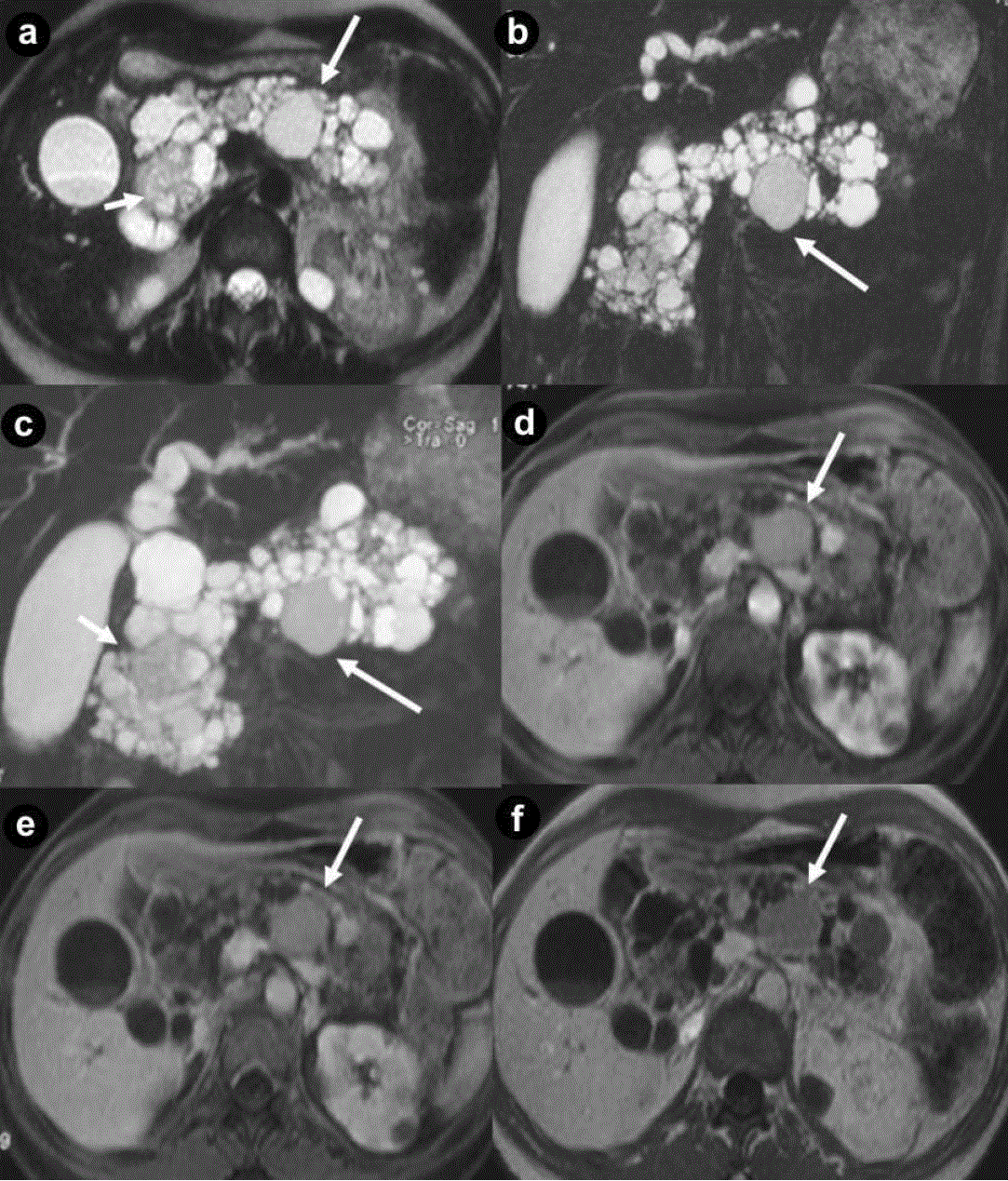
| Figure 5. Pancreatic diffuse microcystic serous cystadenoma and pancreatic non functioning neuroendocrine tumor in the same patient. Asymptomatic 34-year-old woman, member of family affected to VHL disease. Axial (a.) and coronal (b.) T2-weighted MR images, MR cholangiopancreatography (c.) and axial 3D volumetric gradient-echo T1-weighted fat suppressed images after intravenous contrast medium administration during arterial pancreatic (d.), portal venous (e.) and delayed (f.) phases of contrastographic dynamic study. Pancreatic gland is enlarged and parenchyma is completely replaced by multiple, lobulated fluid cysts, hyperintense on T2-weighted MR images, with a “bunch of grapes” pattern (a. b. c.). The wall of cystic lesions and septa inside them shows enhancement after intravenous contrast medium administration during arterial pancreatic (d.), portal venous (e.) and delayed (f.) phases. In pancreatic head and neck, some cysts result so small and so numerous as they show lower signal intensity on T2-weighted MR images compare to fluid lesions and appear at MR imaging like solid mass (short arrow). In the pancreatic body, a round solid mass (arrows), with the signal intensity less high than the other pancreatic cysts on T2-weighted MR images and homogeneous enhancement after intravenous contrast medium administration during arterial pancreatic phase (d.) with low wash-out during portal venous phase (e.) but hypointense on delayed (f.) phase, is present. Multiple bilateral renal fluid cysts are detected. Splenopancreatectomy was performed. Histological specimen showed the presence of pancreatic diffuse serous cystadenoma and non functioning neuroendocrine tumors of pancreatic body-tail. |