|
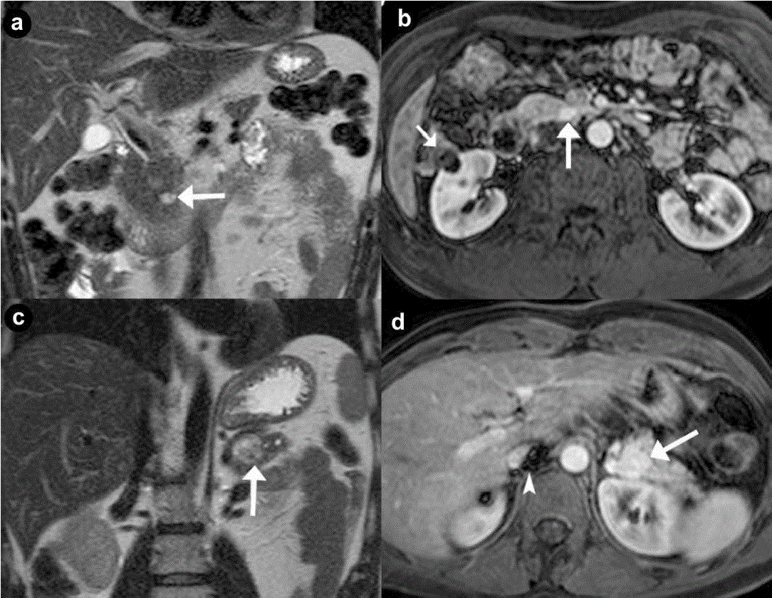
| Figure 2. Pancreatic non functioning neuroendocrine tumors. Two cases suffering from VHL syndrome: a 39-year-old man (a. b.), and a 37- year-old man (c. d.). Coronal (a. c.) T2-weighted MR images; axial (b. d.) 3D volumetric gradient-echo T1-weighted fat suppressed images after intravenous contrast medium administration during arterial pancreatic phase of contrastographic dynamic study. In pancreatic head, a small, nodular solid lesion with maximum diameter of 12 mm (a. b. arrow), hyperintense on T2-weighted MR images, with homogeneous enhancement after intravenous contrast medium administration during arterial pancreatic phase is present. Main pancreatic duct is normal and common bile duct is not dilated. Small complex cystic mass in the right kidney without enhancement after intravenous contrast medium administration (b.) is present (short arrow). In pancreatic body-tail a solid, round mass with maximum diameter of 23 mm (c. d. arrow), heterogeneously hyperintense on T2-weighted MR images, with homogeneous enhancement after intravenous contrast medium administration during arterial pancreatic phase, is present. Artifacts are recognizable in the site of right adrenal gland (arrowhead), due to the presence of metallic staples of previous adrenalectomy for pheochromocytoma. |