|
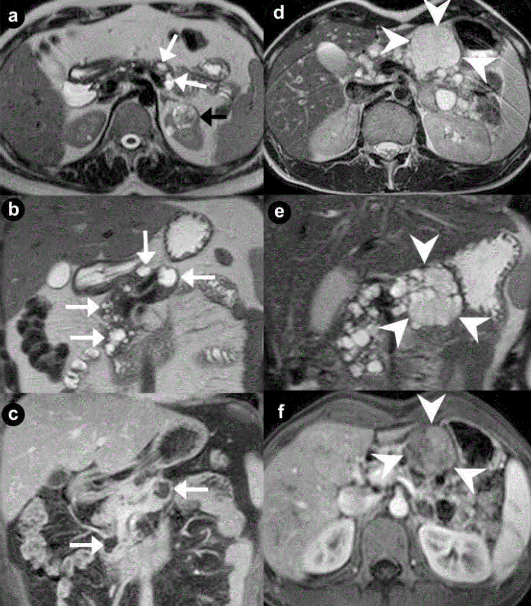
| Figure 1. Pancreatic unilocular cysts (a. b. c. Case#1: 35-year-old man with VHL disease). Pancreatic microcystic serous cystadenoma (d. e. f. Case#2: 30-year-old man with VHL disease). Axial (a. d.) and coronal (b. e.) T2-weighted MR images; coronal (c.) and axial (f.) 3D volumetric gradient-echo T1-weighted fat suppressed images after intravenous contrast medium administration during arterial pancreatic phase of contrastographic dynamic study. Case#1. Multiple fluid, cystic lesions of pancreatic parenchyma with different size and site (arrows), hyperintense on T2-weighted MR images, are present (a. b. c.). Cystic lesions do not communicate with the main pancreatic duct, which appears not dilated. The cystic walls do not show enhancement after intravenous contrast medium administration (c.). In the upper pole of the left kidney (a.) a complex cystic mass (black arrow) is detected. Case#2. A lobulated fluid mass, with thin walls, hyperintense at T2-weighted images (d. e.), is present in the body of pancreatic gland (arrowheads). Inside the mass, containing multiple fluid cystic areas, multiple radially aligned thin septa are visible (spongy appearance or honeycomb pattern). The septa and the peripheral walls enhance after intravenous gadolinium administration (f.). The septa are well depicted on coronal T2-weighted MR image (e.) but the central scar is not visible. In the remaining portion of pancreatic gland multiple cystic, fluid, round lesions, with different size and site and without enhancement after intravenous contrast medium administration (f.), are present. |