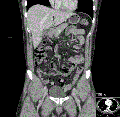
Figure 2.
A computed tomography scan showed a heterogeneous 4 cm malignant-looking lesion in the gastric antrum with suspected invasion of the perigastric fat tissue.