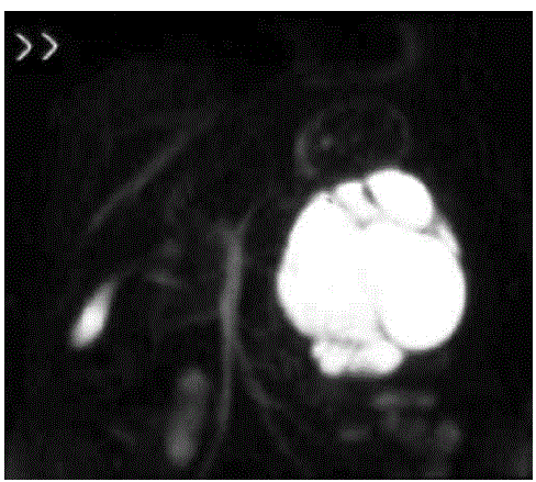
Figure 5:
Coronal FSE/SPIR of mucinous pancreatic neoplasm (peripheral type). MR image shows lobulated, macrocystic pancreatic mass with cystic components located in the tail