|
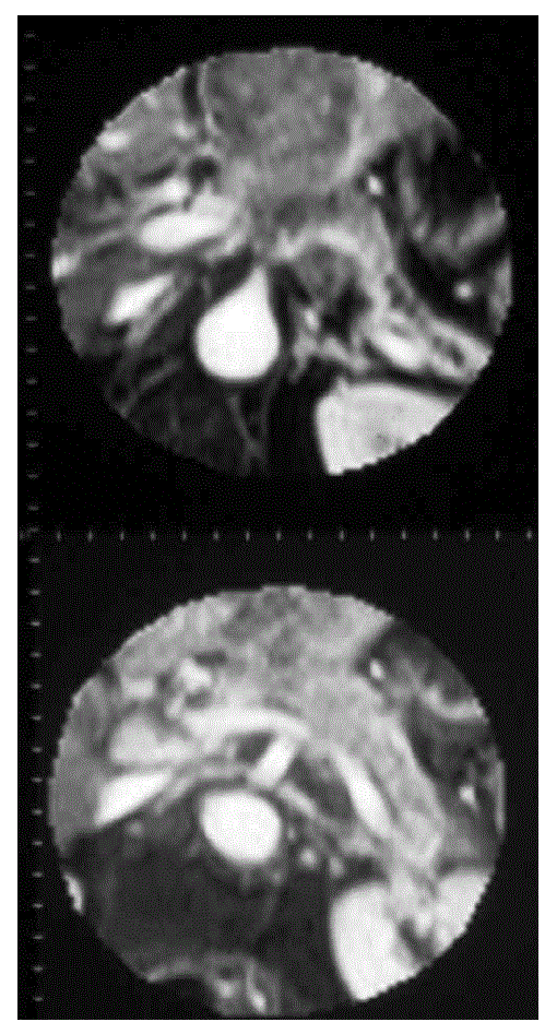
| Figure 4:Contrast-enhanced RF fast T1-weighted MR images (same case) show pancreatic neoplasm infiltrating the hepatic and splenic arteries at the origin (upper) and the splenic vein (lower). Slight dilatation of the main pancreatic duct is also visible (upper). |