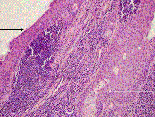
Figure 3.
Photomicrograph of H&E stain at 20x magnification showing stratified squamous epithelium (black arrow), sebaceous units (white arrow), and surrounding lymphocyte infiltration.